Imaging Medical Devices Market: Revolutionary Technologies Redefining Global Healthcare Delivery
So, what is medical imaging exactly? At its core, medical imaging encompasses the diagnostic technologies that allow healthcare providers to peer inside the human body without making a single incision. Think of it as giving doctors a window into what’s happening beneath the skin, enabling accurate disease detection and monitoring.
The imaging medical devices market hit an impressive $18.7 billion in valuation during 2026, and based on our research, it’s projected to expand toward $28.2 billion by 2034. That’s a solid 5.23% compound annual growth rate throughout the forecast period.
What Are the 4 Types of Medical Imaging Devices?
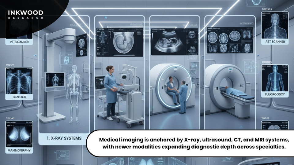
When people ask about the four primary types of medical imaging devices, they’re usually referring to the workhorses of diagnostic healthcare.
- X-ray systems, the oldest and most widely recognized imaging modality, have been around for over a century and remain essential for everything from broken bones to dental checkups.
- Ultrasound equipment takes a completely different approach by using sound waves to generate real-time images and it’s the same technology that lets expecting parents see their babies before birth.
- Computed tomography (CT) scanners take things up a notch by combining multiple X-ray measurements to create detailed cross-sectional views of the body.
- Meanwhile, magnetic resonance imaging (MRI) systems use those uniform magnetic fields created by advanced medical imaging devices to produce incredibly detailed soft tissue images that other modalities simply can’t match.
However, here’s where it gets interesting: diagnostic imaging medical devices actually extend far beyond these four fundamental categories:
- Positron emission tomography (PET) scanners, for instance, detect metabolic activity by tracking radioactive tracers moving through the body.
- Mammography systems have been specifically designed for breast cancer screening, while nuclear imaging equipment offers unique insights by monitoring radioactive materials within tissues.
- Fluoroscopy provides real-time moving images during procedures, allowing physicians to watch as they work.
There’s even a specialized setup called a hybrid operating room; it is an operation theatre equipped with advanced medical imaging devices that seamlessly combines surgical capabilities with sophisticated imaging technologies.
How Does Medical Imaging Work?
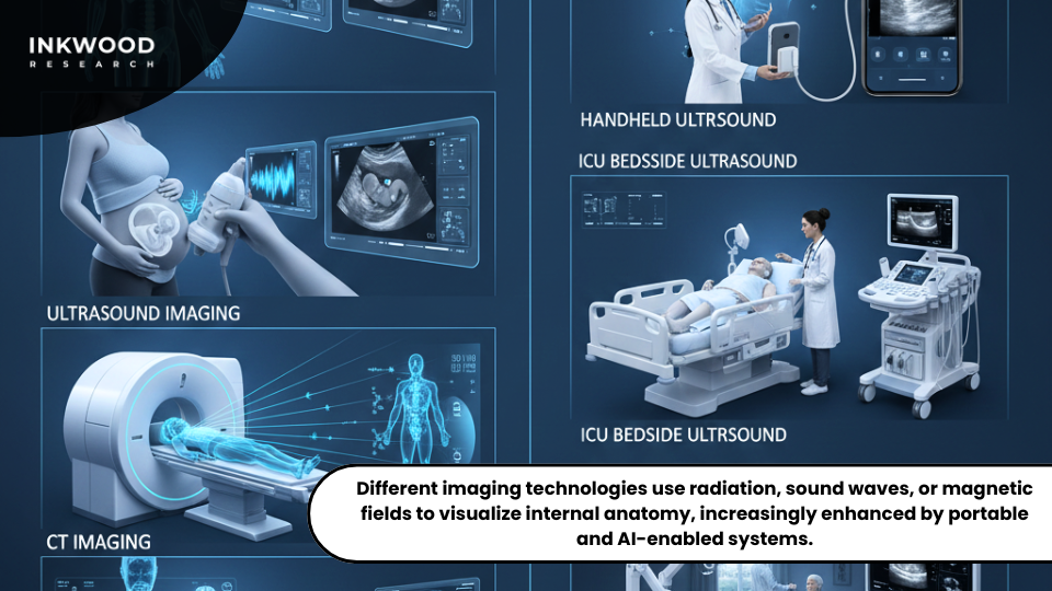
The beauty of medical imaging lies in how different technologies harness various physical principles to reveal what’s hidden inside us. X-ray systems work by passing electromagnetic radiation through the body, where dense tissues like bones absorb more radiation than soft structures, creating those distinctive skeletal images we all recognize.
Ultrasound devices take a gentler approach, transmitting high-frequency sound waves that bounce off internal organs and return as echoes, which the machine then converts into real-time images. CT scanners add complexity by rotating X-ray sources around patients, capturing images from multiple angles and reconstructing them into detailed three-dimensional anatomical maps.
MRI machines might seem like magic, but they’re actually manipulating hydrogen atoms throughout your body using powerful magnetic fields and radio waves, generating exceptional tissue differentiation that helps doctors spot issues other technologies might miss.
What’s truly exciting is how portable medical imaging devices now deliver diagnostic quality that rivals traditional stationary equipment. Battery-powered ultrasound units let doctors perform bedside examinations in emergency rooms or intensive care units, eliminating the need to transport critically ill patients.
Mobile X-ray systems can reach homebound patients who simply can’t make it to imaging centers. Among the most innovative imaging devices examples are pocket-sized ultrasound scanners that connect directly to smartphones, essentially putting diagnostic capabilities in physicians’ pockets.
These technological breakthroughs are democratizing healthcare access in ways that would have seemed impossible just a decade ago, reaching across geographic and economic barriers that previously limited who could access quality diagnostic care.
Poland's Government-Funded AI Revolution in Mobile Imaging
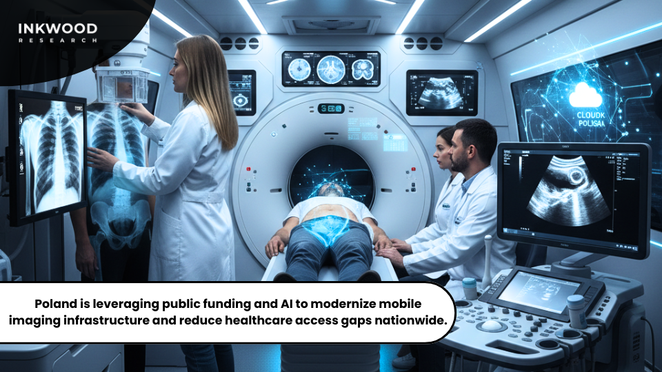
Something remarkable is happening in European healthcare, and Poland is leading the charge with strategic government investments in artificial intelligence capabilities. The Poland mobile imaging services market hit $190.47 million in 2026 and is expected to climb toward $243.24 million by 2034—a steady 3.10% compound annual rate that tells only part of the story.
What makes this particularly interesting is how Poland is using targeted public funding to modernize medical technology infrastructure in ways that could become a blueprint for other nations.
Strategic Government Investment Framework
Poland’s National Reconstruction Plan isn’t playing around. They’ve earmarked approximately $400 million by 2026 specifically for AI-driven medical technologies. This isn’t just throwing money at the problem; it’s a focused investment directly supporting diagnostic imaging infrastructure expansion across both bustling urban centers and historically underserved rural communities.
Healthcare institutions throughout the country are leveraging these governmental funds to acquire cutting-edge mobile imaging equipment that incorporates AI algorithms for enhanced diagnostic accuracy, fundamentally changing how healthcare reaches Polish citizens. Mobile units equipped with digital radiography, computed tomography, and ultrasound capabilities are increasingly becoming a common sight serving Polish populations that previously had to travel hours for basic diagnostic services.
These strategic investments address geographic healthcare disparities head-on while simultaneously improving overall population health outcomes. The PIAST-AI Factory initiative takes things even further by integrating high-performance computing resources with cloud-based AI platforms specifically designed to serve healthcare and life sciences sectors, creating an ecosystem where innovation can thrive.
AI Integration Transforms Diagnostic Workflows
Here’s where things get really interesting. Artificial intelligence applications are revolutionizing how Polish healthcare providers deliver mobile imaging services in practical, everyday ways.
- Machine learning algorithms are slashing image interpretation times while actually improving radiologist accuracy in detecting those subtle abnormalities that might otherwise slip through.
- Cloud-based enterprise imaging solutions now enable seamless data integration across distributed health systems, meaning a patient’s imaging history can follow them wherever they receive care.
- Real-time AI assistance even supports technicians during image acquisition, automatically optimizing positioning and exposure parameters without requiring manual adjustments.
As a result, mobile imaging providers that incorporate AI capabilities are gaining substantial competitive advantages in both diagnostic accuracy and operational efficiency. Workflow automation cuts per-examination costs while simultaneously increasing how many patients can be seen each day. This efficiency improvement proves particularly valuable given Poland’s rising chronic disease prevalence, which continues driving sustained demand for diagnostic imaging services.
The Polish mobile imaging market demonstrates something important; when you combine targeted government funding with genuine technological innovation, you create sustainable healthcare infrastructure improvements that benefit entire populations rather than just privileged segments. This European model offers valuable lessons as neighboring nations wrestle with similar healthcare modernization challenges and demographic pressures.
Medicare 2026 Fee Schedule Reshapes U.S. Mobile Imaging Economics
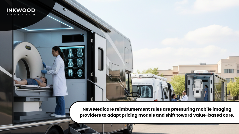
American healthcare providers are navigating some pretty significant changes when it comes to reimbursement for mobile medical imaging services, and these shifts are creating real challenges for businesses trying to plan sustainable operations.
The Centers for Medicare & Medicaid Services dropped their finalized 2026 Medicare Physician Fee Schedule, introducing dual conversion factors that substantially impact how diagnostic imaging gets reimbursed. If you’re running a mobile imaging operation or thinking about starting one, understanding these regulatory shifts is essential for survival.
Dual Conversion Factor Implementation
Starting in January 2026, Medicare is doing something it’s never done before: implementing separate conversion factors that distinguish qualifying Alternative Payment Model participants from everyone else. If you’re a qualifying APM participant, you’ll see a conversion factor of $33.57, which represents a 3.77% increase over 2025 levels. Not terrible, right?
However, non-APM participants face a lower conversion factor of $33.40, translating to just a 3.26% increase. This bifurcated structure is clearly designed to push imaging providers toward value-based care arrangements if they want optimal reimbursement rates. But here’s the kicker that has radiologists up in arms.
Both conversion factors incorporate a controversial -2.5% efficiency adjustment that specifically targets non-time-based services, including diagnostic imaging interpretation. The Centers for Medicare & Medicaid Services maintains that technological advancements and standardized workflows have reduced the resources these procedures require, so reimbursement should reflect that reality.
The result? Radiology is looking at an estimated -2% overall impact, while nuclear medicine faces -1% reimbursement pressure under the finalized fee schedule changes. Industry groups have fought hard against this adjustment, arguing it doesn’t reflect actual practice costs, but CMS wasn’t swayed.
Mobile Imaging Service Implications
These Medicare policy modifications are creating some genuinely complex financial planning headaches for mobile medical imaging businesses across the United States. Operators of portable medical imaging devices need to completely recalculate their service pricing structures to account for these reduced reimbursement rates, which isn’t as simple as just adjusting a few numbers in a spreadsheet.
Besides, American reimbursement policies don’t just affect U.S. providers—they influence equipment purchasing decisions worldwide as manufacturers design products to meet stringent Medicare requirements.
Indian entrepreneurs launching mobile X-ray businesses study U.S. regulatory precedents when structuring their own service delivery models. Medicare policy essentially creates ripples that extend far beyond American borders, shaping international medical imaging equipment specifications and operational protocols that get adopted globally.
Launching Mobile X-Ray Ventures in India's Expanding Market
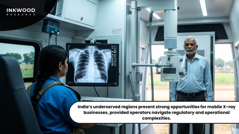
Entrepreneurial healthcare providers are recognizing some substantial opportunities in establishing mobile X-ray businesses throughout India’s vast and underserved population. The subcontinent’s healthcare infrastructure has significant gaps that create nearly perfect conditions for deploying portable medical imaging devices to reach millions who currently lack convenient access to diagnostic services.
Starting a mobile radiology venture isn’t exactly straightforward, though. It requires careful planning around regulatory compliance, equipment acquisition, and building a sustainable business model that can actually survive in competitive markets.
Essential Startup Requirements
If you’re wondering how to start a mobile X-ray business in India in 2026, the journey begins with comprehensive market research to identify underserved geographic regions that actually need your services. Rural areas offer particularly attractive opportunities since populations there often lack nearby diagnostic facilities, but you can’t just set up shop anywhere.
Initially, you’ll need to choose between operating as a sole proprietorship, forming a partnership, or establishing a limited liability company. Each structure carries distinct tax implications and liability protections that can dramatically affect your financial outcomes and personal risk exposure.
India’s healthcare regulatory framework also requires specific licenses and permits that vary depending on which state you’re operating in, adding layers of complexity. Registration with medical councils and radiation safety authorities represents mandatory compliance steps before you can legally commence operations. Well, skip these at your peril.
Additionally, aspiring operators should absolutely prioritize reputable suppliers who offer comprehensive warranties and 24/7 technical support, because when your equipment goes down in the field, you’re losing money by the hour. Equipment reliability directly impacts your business reputation and customer satisfaction in markets where word-of-mouth can make or break your success.
Cardiac MRI Excellence in Australia and New Zealand
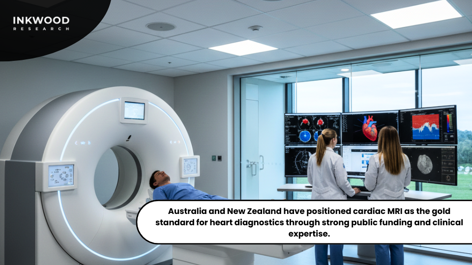
Cardiovascular imaging stands as one of the most technically demanding applications across the entire medical imaging technologies spectrum. Accordingly, Australia and New Zealand have established themselves as genuine leaders in this specialized field.
These nations have embraced cardiac magnetic resonance imaging as the undisputed gold standard for comprehensive heart disease evaluation, and their healthcare systems prioritize advanced diagnostic capabilities that consistently deliver superior patient outcomes through evidence-based technology adoption.
Why Cardiac MRI Achieves Gold Standard Status
Cardiac MRI in 2026 delivers soft tissue contrast and functional assessment capabilities that alternative imaging modalities simply can’t match. Unlike computed tomography which requires radiation exposure and contrast agents that some patients can’t tolerate, MRI harnesses those uniform magnetic fields created by advanced medical imaging devices to produce remarkably detailed images without any ionizing radiation whatsoever.
This safety advantage proves particularly valuable for young patients who need their hearts monitored over decades, as well as those requiring repeated follow-up examinations throughout chronic disease management where cumulative radiation exposure becomes a legitimate concern.
What makes cardiac MRI truly impressive is how it provides comprehensive anatomical and functional information within single examination sessions that would otherwise require multiple separate tests. Cardiologists can assess ventricular volumes, ejection fractions, myocardial viability, and perfusion dynamics all at once, painting a complete picture of heart health in ways that piecemeal testing never could.
Healthcare System Support Framework
Australia and New Zealand’s universal healthcare systems facilitate broader cardiac MRI access compared to fee-for-service healthcare models that dominate in places like the United States.
- Government funding supports academic medical centers that maintain specialized cardiac imaging expertise and the expensive equipment infrastructure required for high-quality programs.
- Public hospitals offer cardiac MRI services to referred patients without any direct out-of-pocket costs, creating genuinely equitable access across socioeconomic strata rather than limiting advanced diagnostics to the wealthy.
That said, no system is perfect, and demand occasionally outstrips available capacity, resulting in examination wait times that can extend several weeks for non-urgent cases.
- Private imaging centers have stepped in to supplement public sector capacity for patients willing to pay out-of-pocket costs or those who possess private health insurance coverage.
This mixed public-private model attempts to balance universal access principles with the capacity constraints that affect virtually all publicly funded healthcare systems globally, though reasonable people disagree about whether the balance is struck correctly.
Australian and New Zealand cardiologists also benefit from robust professional education programs that support advanced imaging interpretation skills you don’t develop overnight. Professional societies conduct regular training programs ensuring practitioners actually maintain current knowledge of evolving cardiac MRI protocols and applications rather than relying on outdated training from years past.
Public Versus Private MRI Access in Canada
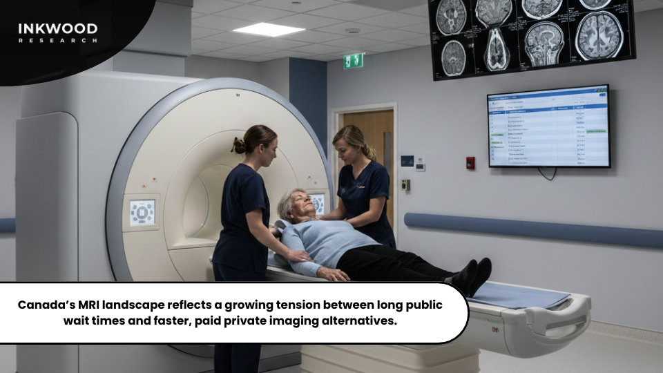
Canadian healthcare consumers are increasingly facing some tough choices between waiting months for publicly funded MRI access or shelling out their own money for private clinic services that can get them scanned within days. Read more about it here.
This tension reflects much broader debates about healthcare equity, resource allocation, and how to sustainably finance universal healthcare systems that actually work. Understanding these trade-offs helps both patients making personal decisions and policymakers trying to navigate the complex balance between cost, access, and quality considerations.
Public System Wait Time Challenges
Canada’s publicly funded healthcare model provides MRI examinations without charging patients directly for medically necessary imaging; a principle Canadians fiercely defend as fundamental to their national identity. However, capacity constraints create significant wait times that vary substantially across provinces and depend heavily on how urgent your case is deemed.
Non-urgent diagnostic imaging requests may face delays extending several months in some jurisdictions, which sounds reasonable on paper until you’re the one living with unexplained symptoms wondering if something serious is being missed. These wait times generate understandable patient frustration and can have real clinical consequences when diagnostic delays impact treatment planning or allow diseases to progress unchecked.
Provincial governments continuously work on improving imaging capacity through equipment purchases and technologist recruitment, throwing money at the problem in hopes it’ll eventually catch up. Nevertheless, demand growth driven by aging populations and expanding clinical indications perpetually outstrips available resources, like trying to bail out a boat with a leaky bottom.
Budget limitations restrict how quickly capacity can expand despite those growing wait lists that keep politicians up at night. As a result, patients with financial means are increasingly seeking private alternatives that let them circumvent public system delays entirely, creating a two-tier system that makes equity advocates deeply uncomfortable.
Private Imaging Center Economics
Private MRI clinics operating across Canada offer dramatically reduced wait times, typically providing examinations within days or weeks of referral rather than the months you might wait in the public system. Patients pay directly for these services out of pocket, with costs ranging from $600 to $1,200 CAD depending on examination complexity and which part of your body needs scanning.
Critics argue that private imaging actually exacerbates inequalities by allowing wealthy Canadians to bypass public wait times while middle and lower-income populations remain stuck waiting, constrained by capacity limitations they can’t pay their way around. Supporters counter that private services actually reduce the burden on public systems by diverting patients willing to pay privately, potentially shortening wait times for everyone else who relies exclusively on public access—though the data on whether this actually happens remains hotly contested.
The question “Is paying for faster scans worth it?” honestly lacks simple answers since it depends so much on your individual circumstances and clinical context. Patients with acute symptoms requiring urgent diagnosis may find private imaging absolutely invaluable in preventing disease progression during those crucial wait periods.
Others dealing with stable chronic conditions may reasonably decide they can accept longer wait times to avoid spending money they’d rather use elsewhere. Canadian MRI access debates ultimately illuminate fundamental tensions within universal healthcare systems worldwide—how do you balance equity principles against capacity realities and respect for individual choice?
These challenges connect directly to very different discussions emerging in the UAE, where pediatric imaging access gets addressed through specialized service design enabled by entirely different healthcare financing models.
Child-Friendly Pediatric MRI Solutions in the UAE
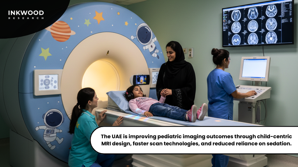
Pediatric medical imaging presents unique technical and psychological challenges requiring specialized equipment and protocols addressing children’s developmental needs. The United Arab Emirates healthcare sector invested substantially in child-friendly imaging solutions recognizing that standard adult-oriented facilities often traumatize young patients. These innovations demonstrate how thoughtful facility design and technology adaptation improve diagnostic quality while enhancing patient experiences.
Pediatric Imaging Challenges
Young children struggle remaining motionless during MRI examinations typically requiring 20-45 minutes of complete stillness. Movement artifacts degrade image quality necessitating examination repetition exposing children to prolonged scanner time. Additionally, MRI machines’ confined spaces and loud operational noises frighten pediatric patients unaccustomed to clinical environments.
Consequently, many facilities historically relied on sedation or general anesthesia enabling examination completion, introducing additional risks and costs. Furthermore, children’s smaller anatomical structures require specialized imaging coils and protocol adjustments optimizing signal-to-noise ratios for diagnostic quality images.
Standard adult protocols often produce suboptimal pediatric images limiting diagnostic confidence. Radiation considerations prove particularly important for X-ray and CT examinations given children’s greater lifetime cancer risk from ionizing radiation exposure.
UAE’s Child-Friendly Design Innovations
Pediatric MRI in the UAE incorporates environmental design elements reducing anxiety and improving cooperation rates. Imaging suites feature colorful decorations, character themes, and entertainment options creating welcoming atmospheres rather than intimidating clinical spaces.
Wide-bore MRI scanners provide less claustrophobic environments accommodating anxious children more comfortably. Some facilities install audiovisual systems allowing children watching movies during examinations distracting from scanner noises. Additionally, child life specialists trained in pediatric psychology work with young patients preparing them for imaging experiences through age-appropriate explanations and practice sessions.
Mock MRI scanners allow children familiarizing themselves with equipment and sounds before actual examinations. Parents often remain present during procedures providing emotional support reducing anxiety levels. Moreover, technological advances enable faster imaging sequences reducing total examination times.
Compressed sensing techniques and parallel imaging algorithms maintain diagnostic quality while cutting scan durations significantly. Shorter examinations improve pediatric cooperation rates reducing sedation requirements and associated risks. These innovations reflect broader medical imaging technologies evolution prioritizing patient-centered care delivery models.
Leading Medical Display Monitor Manufacturers in America
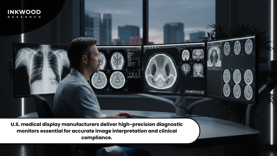
Medical display monitors constitute essential components of modern diagnostic imaging workflows enabling radiologists interpreting images with precision and accuracy. The United States market features numerous specialized manufacturers producing displays meeting stringent regulatory requirements for clinical diagnostic applications. Understanding top medical display monitor brands helps healthcare facilities making informed purchasing decisions balancing image quality, reliability, and cost considerations.
Top 10 Medical Display Monitor Brands
- Barco dominates the premium medical display segment with diagnostic monitors featuring exceptional brightness uniformity and grayscale performance. The Belgian manufacturer’s displays incorporate automatic calibration systems maintaining consistent image quality throughout product lifecycles.
- Sony Healthcare Solutions produces high-resolution surgical and diagnostic displays leveraging proprietary imaging technologies developed through consumer electronics expertise.
- EIZO Corporation specializes in medical imaging displays with comprehensive quality assurance software and extended warranties addressing healthcare reliability requirements.
- LG Electronics entered medical display markets offering competitively priced diagnostic monitors challenging traditional premium manufacturers.
- Dell Technologies provides enterprise-grade displays suitable for non-diagnostic clinical review applications at attractive price points.
- JUSHA Medical manufactures comprehensive display portfolios serving diverse clinical specialties from radiology to surgical visualization.
- NDS Surgical Imaging focuses on operating room displays designed for hybrid operating theaters integrating imaging medical devices with surgical workflows.
- Furthermore, FSN Medical Technologies specializes in modular display solutions adaptable to evolving clinical requirements.
- Advantech produces medical-grade computers and displays emphasizing infection control features and fanless cooling designs.
- Quest International delivers custom display solutions for specific clinical applications requiring specialized configurations.
These manufacturers collectively address diverse healthcare facility needs spanning academic medical centers to small outpatient imaging clinics.
Display Technology Specifications
Medical display monitors differ substantially from consumer displays through stringent performance specifications and regulatory compliance requirements. Luminance consistency across display surfaces ensures uniform brightness preventing diagnostic errors from uneven image presentation.
Grayscale calibration maintains precise tonal reproduction critical for detecting subtle pathology. DICOM calibration standards define specific luminance curves optimized for medical image interpretation. Additionally, medical displays incorporate quality assurance software continuously monitoring performance parameters. Automated alerts notify technical staff when displays drift outside acceptable performance ranges requiring recalibration or replacement.
These quality control systems prove essential maintaining accreditation compliance and medicolegal risk management. Resolution requirements vary by clinical application, with digital mammography demanding exceptionally high pixel densities detecting microcalcifications indicating early breast cancer.
Moreover, ergonomic considerations influence display selection decisions. Radiologists spend extended periods reviewing images, making display positioning, anti-glare coatings, and blue light reduction features important preventing visual fatigue and musculoskeletal disorders.
Therefore, American healthcare facilities carefully evaluate medical display monitor options balancing technical specifications, regulatory compliance, ergonomic features, and total cost of ownership across expected product lifecycles. These considerations extend globally as Asian markets rapidly expand medical display technology adoption driven by healthcare infrastructure investments and growing diagnostic imaging volumes.
Asia's Accelerating Medical Display Technology Integration
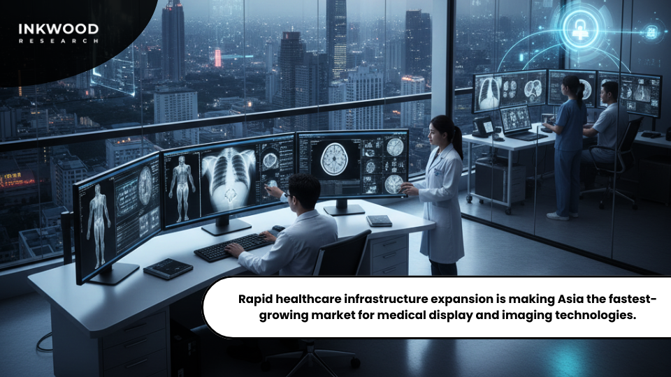
Regional healthcare technology adoption patterns reveal Asia emerging as the fastest-growing market for medical display monitors and associated diagnostic imaging infrastructure. Economic development, healthcare spending increases, and government policy initiatives converge creating unprecedented opportunities for medical technology suppliers. Understanding these regional dynamics helps manufacturers and healthcare providers capitalizing on explosive growth reshaping global medical imaging devices market geography.
Market Growth Drivers
Asia’s medical display market expansion reflects broader diagnostic imaging infrastructure buildout across developing economies. China’s hospital construction boom drives massive equipment procurement as new facilities require comprehensive imaging departments. India’s expanding private healthcare sector invests heavily in advanced diagnostic capabilities attracting medical tourism and domestic middle-class patients.
Southeast Asian nations modernize aging healthcare infrastructure replacing obsolete equipment with current-generation technologies. Additionally, government initiatives prioritize healthcare accessibility improvements across underserved rural populations. Telemedicine programs connecting rural clinics with urban specialty centers require high-quality displays enabling remote diagnostic consultation.
Mobile health units equipped with portable medical imaging devices need ruggedized displays functioning reliably under challenging environmental conditions. These applications create diverse display requirements spanning premium diagnostic monitors to cost-optimized solutions suitable for resource-constrained settings.
Japan's Aging Demographics Drive Imaging Technology Demand
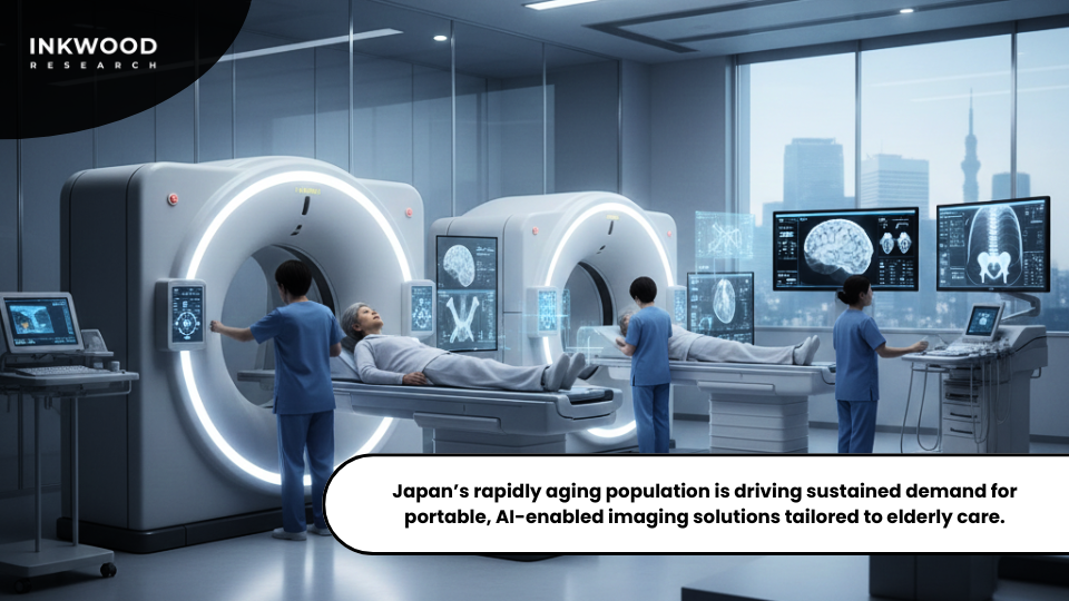
Japan confronts population aging unparalleled among developed economies with profound implications for healthcare delivery systems and medical technology markets. Currently, over 29% of Japanese residents exceed 65 years of age, and projections indicate this proportion reaching 38% by 2060. This demographic transformation creates sustained demand for diagnostic imaging services as elderly populations experience higher disease incidence requiring frequent medical evaluation and monitoring.
Aging Population Healthcare Implications
Elderly patients undergo substantially more imaging examinations than younger demographics due to chronic disease prevalence increasing with age. Cardiovascular conditions, cancer, osteoporosis, and neurodegenerative diseases all require diagnostic imaging for detection, staging, and treatment monitoring.
Japan’s universal healthcare coverage ensures elderly citizens access necessary diagnostic services without financial barriers, supporting consistent imaging utilization regardless of economic circumstances. Moreover, Japan’s aging population influences specific imaging technology adoption patterns. Portable and AI-powered imaging systems gain prominence addressing mobility limitations common among elderly patients.
Bedside ultrasound examinations reduce patient transport requirements within hospitals and nursing facilities. Mobile X-ray services reach homebound elderly individuals unable traveling to imaging centers. These portable medical imaging devices improve care quality while reducing healthcare delivery costs through operational efficiency gains.
Indonesia's AI-Integrated Diagnostic Display Innovation
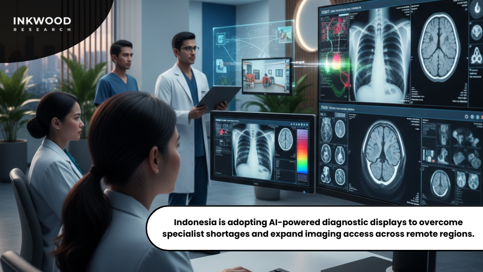
Southeast Asia’s most populous nation emerges as a promising market for next-generation medical display technologies incorporating artificial intelligence and advanced color diagnostic capabilities.
Indonesia’s healthcare system modernization accelerates through both public investment and private sector growth creating opportunities for technology suppliers targeting developing markets. Understanding Indonesia’s unique requirements helps manufacturers positioning products effectively within this high-potential region.
AI-Integrated Color Diagnostic Monitors
The future of medical displays in Indonesia increasingly incorporates artificial intelligence assistance addressing radiologist workforce limitations. AI algorithms embedded within display systems automatically detect abnormal findings highlighting suspicious regions for interpreting physicians. These tools prove particularly valuable in resource-constrained settings where less experienced readers may miss subtle pathology.
Color diagnostic monitors enhance anatomical visualization compared to traditional grayscale displays, improving interpretation confidence for certain clinical applications. Furthermore, cloud connectivity enables centralized image storage and distributed interpretation workflows. Indonesian radiologists in Jakarta interpret examinations acquired in remote Sulawesi clinics thousands of kilometers distant.
This model requires displays maintaining color accuracy and spatial resolution across diverse viewing environments and lighting conditions. Calibration systems must accommodate limited technical support availability in remote locations. Moreover, cost constraints remain paramount across Indonesian healthcare markets.
Display manufacturers must balance advanced capabilities against aggressive pricing requirements competitive within developing market contexts. Local assembly operations and component sourcing strategies reduce costs while demonstrating economic commitment to Indonesian markets.
These considerations reflect broader strategic questions facing medical technology suppliers balancing global product standardization against regional customization requirements. Therefore, Indonesia represents a microcosm of challenges and opportunities characterizing emerging market medical display adoption globally.
The Future of Medical Imaging: From AI-Powered Retinal Screening to Hybrid Operating Rooms
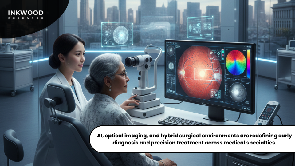
When healthcare systems around the world talk about implementing AI for disease screening, they’re increasingly looking toward South Korea as their model. The nation hasn’t just adopted artificial intelligence for retinal imaging—it’s fundamentally reimagined how early disease detection works, and the results are reshaping ophthalmology across Asia and beyond.
What makes South Korea’s approach particularly innovative isn’t just the technology itself, but how thoroughly it’s been integrated into routine healthcare delivery. AI-assisted diagnostics have moved beyond pilot programs into standard practice at major medical centers, with adoption expanding rapidly into community clinics.
This convergence of ophthalmology with broader systemic medicine represents exactly the kind of innovation that attracted global attention to South Korea’s approach in the first place.
South Korea’s AI Revolution in Retinal Disease Detection
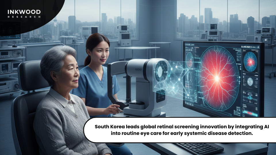
Walk into any major university hospital in Seoul today, and you’ll notice something different about how eye examinations happen. Where traditional fundus photography once required extensive manual interpretation and often caught diseases only after significant progression, AI-powered optical coherence tomography now identifies the earliest cellular changes that precede vision loss.
The numbers tell a compelling story. Diabetic retinopathy affects approximately 15.8% of diabetes patients in South Korea, with vision-threatening cases reaching 4.6% of that population. Yet historically, only about 30% of diabetic patients received the recommended annual dilated fundus examinations. This screening gap existed not because doctors didn’t recognize its importance, but because the traditional examination process created barriers patients found difficult to overcome.
Pupil dilation takes time, causes temporary vision impairment, and requires specialist availability that many regional clinics simply couldn’t provide. AI fundamentally changed this equation. Modern ultra-widefield fundus cameras now capture comprehensive retinal views up to 200 degrees without requiring pupil dilation, making screenings more convenient and accessible than ever before.
Healthcare administrators project that within the next three to five years, AI-powered retinal analysis will become standard protocol for all at-risk populations. This widespread deployment creates network effects that continuously improve diagnostic accuracy, as algorithms learn from ever-larger datasets representing diverse patient populations and disease presentations. The implications extend far beyond ophthalmology.
Retinal imaging increasingly reveals systemic diseases like cardiovascular conditions, neurological disorders, and kidney disease through subtle vascular and neural changes visible in the eye. South Korean researchers are now training AI systems to detect early signs of cognitive decline, hypertension, and autoimmune diseases through retinal biomarkers—essentially turning routine eye examinations into comprehensive health screenings.
India’s Homegrown Retinal Imaging Champions
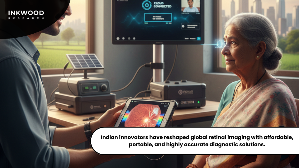
India revolutionized retinal imaging by creating affordable, portable technology for rural healthcare. Unlike expensive tabletop systems requiring dedicated rooms and specialists, Indian companies developed solutions addressing real-world constraints.
- Forus Health pioneered the 3nethra series with handheld retinal cameras capturing high-quality images without pupil dilation. Their cloud-based platforms enable remote diagnosis where specialists don’t exist. Moreover, their AI achieves 95% accuracy for diabetic retinopathy detection, validated across 100,000 patients. With 3,400 installations in 50 countries reaching 10 million lives, Forus competes globally through technological excellence, not just affordability.
- Remidio Innovative Solutions transformed smartphones into diagnostic tools. Their Fundus on Phone device achieves 93-94% sensitivity for detecting diabetic retinopathy—matching traditional cameras at fraction of the cost. FDA clearance validates their technology for U.S. markets.
- Meanwhile, Eyestem Research develops cell replacement therapies like Eyecyte-RPE for age-related macular degeneration, potentially reversing vision loss.
These Indian innovations share a key insight: healthcare technology must address access alongside sophistication. Designing for electricity cuts, limited specialists, and tight budgets created products with universal appeal. Consequently, wealthy healthcare systems now prize the portability, cost-effectiveness, and flexibility Indian manufacturers embedded from inception.
French Ophthalmologists’ Equipment Preferences
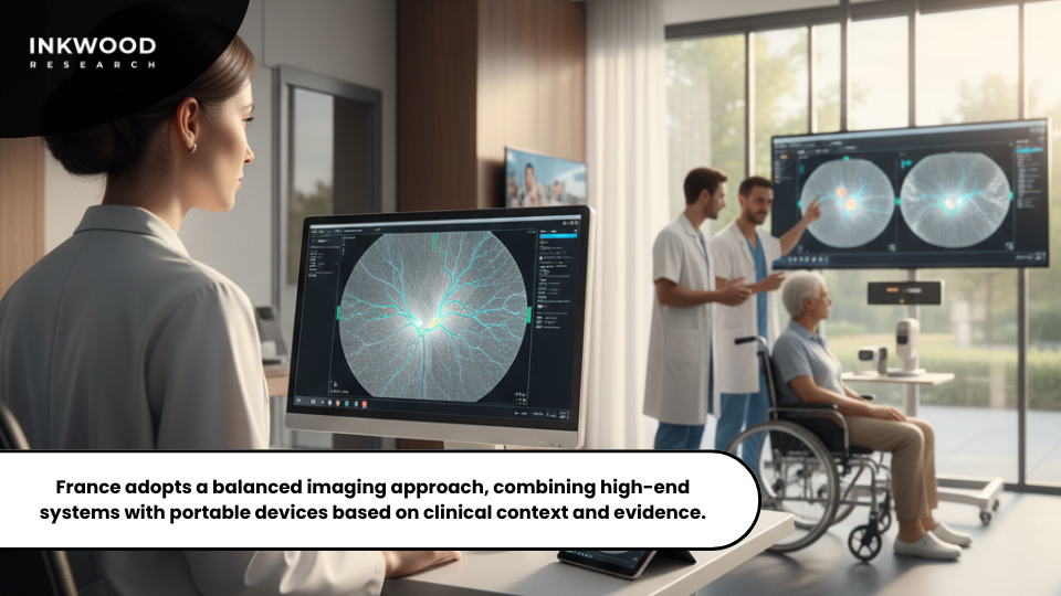
France offers a unique counterpoint to South Korea’s AI-first approach and India’s portability focus. French ophthalmologists, working within well-resourced environments, initially viewed handheld cameras with skepticism. Years using high-end tabletop systems cultivated expectations around image quality that early portable devices couldn’t match.
However, as technology matured, specialists recognized specific clinical advantages. Pediatric ophthalmology benefited significantly, with anxious children tolerating examinations better when devices adapt to them. Similarly, examining patients with mobility limitations became easier with portable equipment.
French academic departments now employ hybrid approaches. They maintain premium tabletop systems for detailed diagnostics while deploying handheld devices for contexts where portability proves decisive. This measured adoption reflects French healthcare culture valuing technological sophistication but demanding rigorous clinical evidence before changing practices.
COVID-19 accelerated telemedicine acceptance. Previously conservative about remote care, French specialists discovered handheld cameras enabled acceptable quality consultations for many conditions. Subsequently, practices found continuing applications: screening programs, satellite clinics, home visits, and collaborative care with optometrists.
France’s evolution illustrates how equipment preferences reflect entire healthcare ecosystems. The “best” retinal camera depends on clinical context, workflow requirements, patient populations, and support infrastructure. French specialists’ thoughtful, evidence-driven adoption provides valuable models for healthcare systems balancing innovation with established practices.
ECG Equipment Market: A Closer look at Inonesia, China, and India
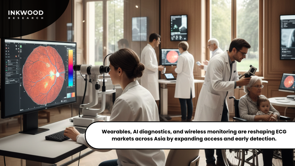
Indonesia reveals how consumer technology disrupts traditional medical device markets. When Apple Watch added ECG capabilities, medical device manufacturers dismissed it as a wellness toy. Indonesian cardiologists shared this skepticism initially.
However, patient behavior proved otherwise. Indonesians arrived at consultations with Apple Watch ECG readings detecting atrial fibrillation episodes that would otherwise cause strokes. Consumer devices weren’t replacing hospital equipment—they expanded the market by identifying previously unknown at-risk patients needing evaluation.
This compelled traditional players like Philips to reconsider strategies. The cardiac monitoring market now bifurcates into diagnostic-grade hospital equipment and continuous monitoring devices for screening. Manufacturers developed wearable solutions while enhancing hospital systems with AI capabilities.
- Wireless ECG technology particularly transforms Indonesia’s geographically dispersed healthcare. Remote patients no longer need transport to Jakarta—wireless devices transmit readings to distant cardiologists for real-time interpretation.
- AI diagnostics detect subtle abnormalities human interpreters might miss. For Indonesia, where cardiologists concentrate in urban centers, AI-assisted interpretation democratizes cardiac care access.
- Wearable devices enable continuous monitoring versus episodic hospital visits, capturing dangerous arrhythmias occurring unpredictably between appointments.
Winning manufacturers recognize cardiac monitoring evolved into an ecosystem requiring different devices for different contexts, including hospital systems, outpatient wireless monitoring, consumer wearables, and AI-powered interpretation software.
China’s Shift to Home-Based Cardiac Monitoring
China’s enormous population and aging demographics drive aggressive shifts toward home cardiac monitoring. With over 290 million people aged 60 or older, traditional hospital-centric models couldn’t accommodate national cardiac monitoring needs. Consequently, necessity drove innovation offering lessons worldwide.
Government policy actively promotes this transition. National healthcare reforms explicitly prioritize chronic disease management at primary care levels. Moreover, reimbursement policies incentivize remote monitoring over inpatient stays. Subsequently, Chinese manufacturers like Lepu Medical, Mindray, and Biolight developed extensive home-use product lines.
Furthermore, China’s approach emphasizes complete ecosystem development rather than individual product excellence. Leading manufacturers provide integrated telemedicine platforms connecting devices, patients, physicians, specialists, and pharmacies. This ecosystem approach acknowledges that home monitoring delivers value only when embedded within workflows using generated data effectively.
Additionally, integration with national health information platforms represents a uniquely Chinese element. ECG data flows into centralized databases where it’s analyzed alongside other health information for population health management. Therefore, early identification of trends enables public health interventions at unprecedented scales.
China’s ECG equipment transformation impresses not through technology alone but through comprehensive health system adaptation. Indeed, product development, regulatory pathways, reimbursement policies, professional training, and information infrastructure evolved coordinately. Meanwhile, Chinese manufacturers increasingly export globally, bringing competitive pressure to established Western medical device companies through high quality at aggressive prices.
India’s Refurbished ECG Market Expansion
India demonstrates how refurbished medical equipment creates parallel markets addressing healthcare access gaps. The refurbished ECG segment evolved from marginal gray market into legitimate, quality-certified industry that hospital administrators view as strategic purchasing rather than budget compromise.
Indian healthcare providers maintain equipment much longer than Western counterparts. Capital constraints make maximum equipment longevity financial necessity. Refurbishment specialists now operate sophisticated facilities meeting international quality standards. They source end-of-life equipment from developed markets, perform comprehensive reconditioning, and provide warranties comparable to new equipment.
This quality improvement transformed refurbished equipment into legitimate strategy for value-conscious institutions. Tier-II and Tier-III hospitals access premium technology at 40-50% discounts. Government facilities expand cardiac care capacity faster than possible with new systems alone.
Original manufacturers initially viewed refurbishment as threatening new sales. However, they increasingly recognize it extends market reach to segments never purchasing new systems. Some manufacturers now operate certified refurbishment programs capturing additional revenue while maintaining quality control.
Leasing and subscription models add another dimension. Healthcare providers access current-generation technology through monthly payments without capital expenditures. This financial innovation makes cardiac monitoring accessible to smaller providers lacking equipment budgets but possessing operational funds.
Optical Imaging's Diagnostic Revolution: Exploring European Markets
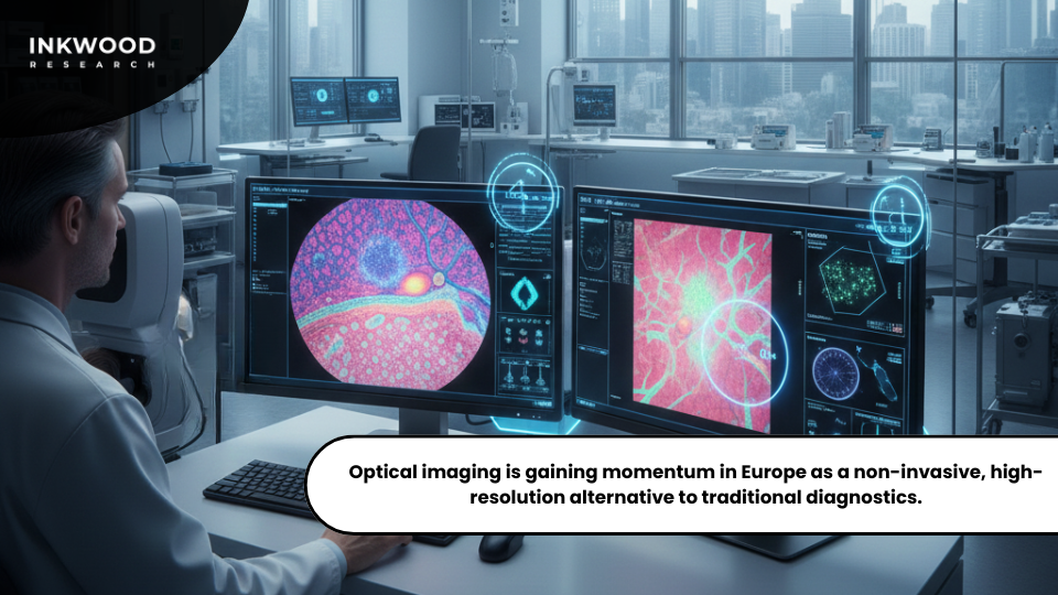
Optical imaging represents a genuinely exciting frontier where technology creates entirely new clinical possibilities. Unlike traditional diagnostics requiring tissue biopsies, optical imaging provides near-cellular resolution non-invasively. This capability transforms workflows across ophthalmology, dermatology, gastroenterology, and oncology.
Moreover, the convergence with artificial intelligence creates powerful multiplier effects. Optical imaging generates massive data volumes containing more information than humans can efficiently process. Consequently, AI algorithms detect subtle early disease patterns, quantify changes with superhuman precision, and guide interventions with unprecedented accuracy.
Clinical applications are expanding rapidly. Ophthalmologists pioneered adoption through optical coherence tomography for retinal disease. Meanwhile, gastroenterologists identify pre-cancerous changes without numerous biopsies. Surgeons now visualize tumor margins during operations using fluorescence techniques that illuminate cancerous tissue.
Intraoperative optical imaging particularly demonstrates transformative potential. Traditional surgery essentially operates blind at microscopic scales. However, optical imaging enables real-time microscopic visualization, allowing surgeons to precisely target disease while confirming complete removal.
Market projections reflect this momentum. Optical imaging represented $3.1 billion globally in 2026, with forecasts projecting growth toward $7.5 billion by 2036. These rates substantially exceed overall medical imaging expansion, reflecting optical imaging’s transition from niche applications toward mainstream adoption across multiple specialties.
Germany’s Optical Imaging Leadership
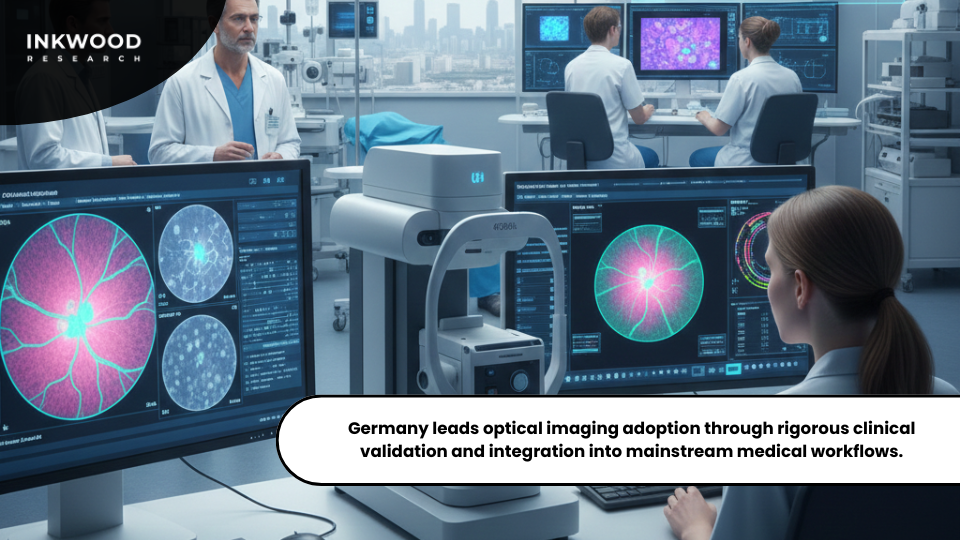
Germany exemplifies how technological sophistication combines with methodical clinical validation. German medical centers lead globally in optical imaging research and adoption, but they always insist on rigorous evidence before widespread deployment.
Germany’s healthcare structure creates ideal conditions for innovation. Strong academic centers maintain advanced research programs. Meanwhile, generous funding supports extended validation studies. Furthermore, comprehensive insurance coverage removes financial barriers once clinical benefits are established.
Optical coherence tomography applications extend well beyond ophthalmology origins. German dermatologists routinely use OCT for non-invasive skin cancer screening, achieving diagnostic accuracies approaching surgical biopsy. Additionally, gastroenterology centers employ optical imaging for early Barrett’s esophagus detection.
The integration into surgical workflows represents particular German strength. Leading centers pioneered fluorescence-guided surgery using molecular probes that illuminate cancerous tissues. Consequently, surgeons visualize tumor margins invisible to the naked eye, ensuring complete removal while minimizing healthy tissue damage.
Dutch AI-Optical Integration
The Netherlands carved a distinctive niche through systematic AI integration with optical imaging hardware. Dutch institutions built perhaps the world’s most sophisticated optical imaging AI ecosystems, combining academic expertise with practical engineering cultures focused on clinically deployable systems.
Collaboration between medical centers, AI institutes, and device companies produced pioneering systems influencing global development. Thirona, a Radboud University spin-off, developed AI platforms emphasizing explainable reasoning rather than inscrutable “black boxes”—critical when clinicians must trust algorithmic interpretations in high-stakes decisions.
Meanwhile, Aidence exemplifies specialized AI addressing clinical bottlenecks. Their Veye Lung Nodule software automatically analyzes chest CT scans, flagging suspicious findings while filtering benign ones. Consequently, this intelligent triaging dramatically improves radiologist productivity.
What makes the Netherlands particularly interesting isn’t individual technologies but the comprehensive ecosystem. The Dutch health system emphasizes data standardization, creating ideal conditions for developing algorithms working across diverse settings. Furthermore, robust privacy frameworks allow extensive clinical data use while protecting patient rights.
Dutch companies have been at the forefront of establishing appropriate AI regulatory oversight frameworks. This regulatory expertise became a competitive advantage, allowing navigation of global approval processes more efficiently than competitors.
Optical Imaging Systems: How South Korea & the US Lead the Market Landscape
South Korea focuses optical imaging development on AI-enhanced early disease detection, representing a strategic priority aligning with strengths in artificial intelligence and preventive healthcare. South Korean researchers systematically applied machine learning to optical imaging data, demonstrating how algorithms identify disease signatures before traditional clinical signs appear.
The most mature applications appear in ophthalmology. Medical centers pioneered AI-powered optical coherence tomography interpretation setting global benchmarks. Moreover, these systems identify subtle structural changes predicting future disease development, enabling interventions before irreversible damage occurs.
South Korea’s retinal imaging success naturally extended to other applications. Dermatology programs now use AI-enhanced confocal microscopy differentiating benign from malignant skin lesions with expert-rivaling accuracy. Meanwhile, gastroenterology centers deploy optical imaging identifying pre-cancerous lesions human endoscopists might miss.
This emphasis on early detection reflects South Korea’s healthcare philosophy prioritizing prevention over treatment. Consequently, optical imaging aligns perfectly with this strategy, offering non-invasive screening across multiple disease categories.
South Korean medical device companies quickly commercialized innovations. Companies like Lutronic developed products specifically optimized for AI-powered screening with interfaces designed for high-throughput workflows. Furthermore, international collaborations amplify impact, with researchers contributing curated datasets from population-wide screening programs, helping train robust algorithms globally.
American Optical Imaging Innovators
The United States approaches optical imaging through characteristic entrepreneurial innovation, with companies developing breakthrough technologies that transform clinical practice in unexpected ways. Top American companies represent fascinating cross-sections of established giants and nimble startups.
- Carl Zeiss Meditec, while German-headquartered, maintains substantial American operations instrumental in bringing optical coherence tomography into mainstream ophthalmology. Their Cirrus HD-OCT platform became the standard, combining exceptional image quality with intuitive workflows. Consequently, advanced imaging became accessible to general ophthalmologists beyond academic specialists.
- Abbott Laboratories entered optical imaging through acquiring OptiMedica, gaining femtosecond laser technologies that transformed cataract surgery. Meanwhile, their integration of OCT into surgical microscopes provides real-time imaging guidance, enabling unprecedented precision.
- Heidelberg Engineering specializes in multi-modal platforms integrating OCT with other modalities. Their approach recognizes no single technique optimally visualizes all clinical features. Therefore, combining modalities provides complementary information improving diagnostic confidence.
- On the startup side, Notal Vision pioneered home-based optical imaging enabling macular degeneration patients to monitor disease progression using smartphone-compatible devices. Furthermore, Optos pioneered ultra-widefield retinal imaging capturing more than 80% of the retina in single images.
What unites successful American companies is problem-focused innovation. They ask “what clinical problems can optical imaging solve better?” rather than “what can we build?”
Dental Support Organizations Transform Equipment Markets
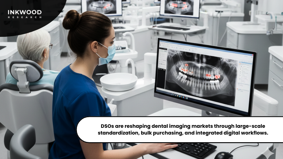
Dental Support Organizations fundamentally transform dental imaging equipment markets by consolidating practices under corporate ownership. DSOs create multi-location networks purchasing equipment at scales individual practitioners could never match. Consequently, this bulk purchasing power changes product requirements, vendor relationships, and innovation priorities.
When DSOs like Heartland Dental standardize imaging equipment across hundreds of locations, that single decision influences markets more than thousands of individual purchases. Therefore, manufacturers increasingly orient strategies toward DSO requirements.
What DSOs demand differs importantly from individual preferences. Standardization across locations matters tremendously—DSOs need equipment dentists can use interchangeably without extensive retraining. Moreover, integration with practice management systems becomes critical at DSO scale. Seamless data flow enables quality monitoring and operational optimization impossible through manual processes.
Service requirements have transformed vendor organizations. Equipment problems across DSO networks create severe aggregated impacts triggering contract penalties. Subsequently, manufacturers must guarantee rapid response across geographic regions.
DSO purchasing cycles also differ dramatically. Individual practitioners purchase opportunistically. However, DSOs make systematic decisions across networks, creating boom-bust cycles for manufacturers.
America’s Dental AI Revolution
Artificial intelligence transforms American dentistry, moving from experimental curiosity to practical tools reshaping daily practice. The United States leads globally in dental AI adoption, driven by technological innovation, regulatory clarity, and market conditions creating strong efficiency incentives.
- Pearl exemplifies how machine learning revolutionizes diagnostic imaging. Their Second Opinion platform analyzes dental X-rays, identifying pathology with expert-matching accuracy. Moreover, clinical studies demonstrate dentists using AI assistance detect 43% more cavities, denoting a stunning improvement translating directly to better outcomes through earlier treatment.
- Meanwhile, Overjet focuses on quantifying disease progression over time. Their AI analyzes sequential X-rays, measuring how cavities enlarge and bone loss advances. Consequently, this enables evidence-based treatment planning with precise measurements rather than subjective impressions.
The FDA’s streamlined approach accelerated American adoption. Most dental AI systems receive Class II classification requiring 510(k) clearance, demonstrating a moderate pathway enabling market entry in 12-18 months rather than many years.
The business case proves compelling. Dentists face pressure improving productivity as reimbursements stagnate while costs rise. Therefore, AI diagnostic assistance genuinely improves per-dentist productivity by helping detect more pathology, make faster decisions, and document findings efficiently.
Japanese Dental Diagnostics Evolution
Japan’s dental AI adoption follows distinctly different patterns from America’s rapid deployment, reflecting cautious innovation culture prioritizing thorough validation. However, dismissing this approach as simply slow would be mistaken as Japanese development emphasizes different priorities that may prove more sustainable.
Japanese dental AI research concentrates heavily on workflow integration rather than standalone diagnostics. Developers recognize isolated diagnostic opinions don’t necessarily improve outcomes if dentists can’t easily incorporate insights. Consequently, Japanese systems embed deeply into practice management platforms, automatically populating treatment plans with AI findings.
The emphasis on patient communication represents another distinctive aspect. Japanese practices value detailed patient education ensuring thorough understanding of proposed treatments. Therefore, AI systems include extensive communication tools—visual aids showing disease progression, comparisons to healthy structures, and projected treatment outcomes.
Major manufacturers like Morita and Yoshida invested heavily in proprietary AI integrated directly into imaging systems. This vertical integration reflects Japanese philosophy that system-level optimization requires controlling all components rather than assembling modular ecosystems.
Hybrid Operating Rooms Across Medical Specialties
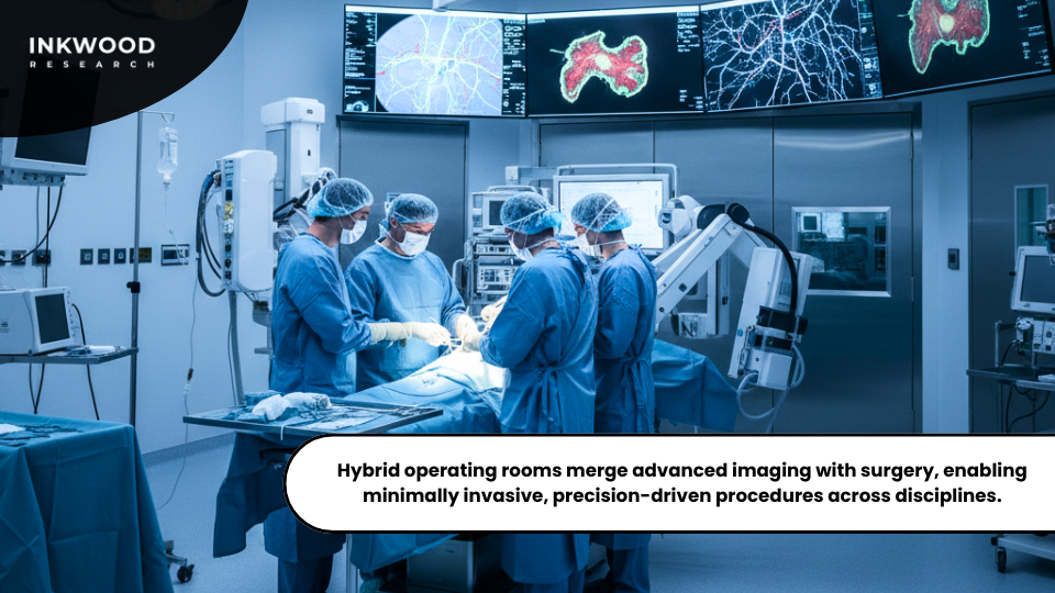
Hybrid operating rooms represent sophisticated medical imaging integration into clinical practice, combining advanced surgical capabilities with comprehensive diagnostic systems providing real-time visualization. While originating in cardiovascular surgery, hybrid ORs now serve diverse specialties discovering unique applications.
Cardiology drove initial development, motivated by minimally invasive cardiac procedures requiring sophisticated imaging. Transcatheter aortic valve replacement exemplifies this—cardiologists perform valve replacements through small arterial punctures using real-time fluoroscopy guidance. Consequently, these procedures demand operating room sterility alongside catheterization laboratory imaging quality.
Neurosurgery applications expanded rapidly as technology matured. Brain tumor resections benefit enormously from intraoperative MRI verifying complete removal before closing incisions. Meanwhile, orthopedic surgery increasingly embraces hybrid capabilities for complex reconstructive procedures requiring precise alignment.
Oncologic surgery represents perhaps the most transformative application. Fluorescence-guided surgery uses specialized imaging visualizing tumor margins invisible to the naked eye. Therefore, surgeons achieve more complete removal while minimizing healthy tissue damage.
What makes hybrid ORs expensive, typically $3-5 million compared to $1-2 million for traditional Ors, is satisfying both surgical sterility requirements and radiation protection standards. Despite costs, adoption accelerates globally as hospitals recognize certain procedures cannot be performed optimally in traditional environments. This favorable economic proposition suggests hybrid ORs will transition toward mainstream infrastructure.
India’s Government-Driven Hybrid OR Growth
India’s hybrid operating room expansion unfolds dramatically differently than developed economies, driven primarily by government initiatives establishing cardiac centers addressing enormous cardiovascular disease burden. This top-down pattern creates unique market dynamics equipment manufacturers must understand.
India’s cardiovascular crisis demands systematic infrastructure responses. Heart disease represents the leading death cause, yet sophisticated care remains concentrated in major urban centers. Therefore, government programs prioritize expanding cardiac capacity in Tier-II and Tier-III cities previously lacking advanced treatment.
These initiatives explicitly mandate hybrid OR capabilities as foundational infrastructure. Modern minimally invasive cardiac procedures require integrated imaging and surgical facilities by definition. Consequently, incorporating hybrid OR requirements ensures facilities deliver contemporary cardiac care.
The financial model differs markedly from developed markets. Indian government facilities evaluate hybrid ORs primarily through population health impact rather than strict financial returns. Subsequently, specifications prioritize cost-effectiveness and reliability over absolute technical sophistication.
Equipment manufacturers must adapt accordingly. Indian buyers value solid clinical performance at reasonable costs more than cutting-edge capabilities commanding premium prices. Moreover, simplified designs reducing maintenance complexity prove particularly attractive given limited biomedical engineering support.
Thailand’s Medical Tourism Hybrid OR Boom
Thailand’s hybrid operating room expansion follows a distinct pattern driven primarily by medical tourism rather than domestic healthcare needs. As medical tourism exploded over the past decade, leading private hospitals invested heavily in hybrid OR capabilities demonstrating technological sophistication to international patients.
Medical tourism reached approximately 2.5 million patients annually before the pandemic, generating over $4 billion in healthcare revenue. This volume concentrates particularly in Bangkok where hospitals compete intensely for patients who carefully research facilities. Therefore, visible technological capabilities like hybrid ORs become important marketing differentiators.
Medical specialties attracting international patients align perfectly with hybrid OR procedures. Cardiovascular interventions, orthopedic surgeries, and oncologic procedures all benefit from hybrid capabilities. Consequently, hospitals like Bumrungrad International and Bangkok Hospital invested in multiple hybrid ORs equipped with advanced imaging systems rivaling American or European medical centers.
Thailand’s medical tourism focus creates interesting ripple effects. The technological sophistication medical tourism demands drives broader healthcare improvements benefiting domestic patients. Moreover, competitive dynamics among hospitals create continuous pressure for technological advancement that government-dominated systems might not sustain.
Regional competition matters enormously. Thailand competes against Singapore, Malaysia, South Korea, and India, carving out positioning as optimal balance of quality and cost. Hybrid OR capabilities communicate this quality message effectively.
German Hybrid Operating Room Market Dynamics
Germany approaches hybrid OR adoption through characteristically systematic planning balancing technological advancement with evidence-based medicine principles and careful cost-effectiveness evaluation. Consequently, this measured approach now positions Germany as a European leader while avoiding less thoughtful implementations characterizing early adoption elsewhere.
Germany’s healthcare structure creates unique conditions for adoption. Strong academic centers maintain research programs rigorously evaluating hybrid OR clinical benefits, generating evidence justifying investments. Moreover, comprehensive insurance coverage removes financial barriers ensuring capacity investments translate into actual patient care.
German hybrid OR development emphasizes comprehensive procedural integration rather than isolated imaging capabilities. Surgical centers redesign entire perioperative workflows to fully exploit hybrid capabilities. Therefore, investments actually change clinical practice rather than adding expensive underutilized capabilities.
Cardiovascular applications have matured most extensively, building on long cardiology excellence. German centers pioneered many complex structural heart procedures now performed worldwide. Furthermore, neurosurgical applications represent another strength, with leading centers developing sophisticated intraoperative imaging protocols maximizing tumor resection while preserving neurological function.
German medical device manufacturers like Siemens Healthineers and KARL STORZ have been instrumental in developing imaging systems and integration platforms. These manufacturers benefit from close clinical collaborations providing rapid feedback, creating innovation ecosystems accelerating technology refinement.
Cardiovascular Ultrasound Market: AI Precision, Asia-Pacific Growth & UAE Opportunity

The cardiovascular ultrasound market is entering a defining decade. Aging populations, rising cardiovascular disease burdens, and a global push toward radiation-free diagnostics are driving strong demand for advanced cardiac imaging solutions.
AI-powered diagnostic precision is reshaping how cardiologists work. Machine learning algorithms now automate left ventricular measurements, wall motion scoring, and strain analysis in real time. Siemens Healthineers’ Acuson Origin system captures over 5,000 cardiac measurements per exam at 99% diagnostic accuracy. GE Healthcare’s 2024 acquisition of Intelligent Ultrasound integrated AI-guided scanning across its portfolio. These advances reduce operator dependency and expand reliable cardiac imaging beyond specialist centers.
Across Asia-Pacific, cardiovascular disease remains the leading cause of mortality. China, India, Japan, and South Korea are all scaling healthcare infrastructure rapidly. Mindray is emerging as a serious regional competitor, offering contrast-capable cardiac platforms at accessible price points across Southeast Asia and beyond.
The UAE cardiovascular ultrasound market reflects the Gulf’s broader healthcare ambition. Government investment through Vision 2031 frameworks is driving hospital capacity expansion across Abu Dhabi and Dubai. Private hospital groups are actively adopting premium AI-enhanced echocardiography systems to serve a growing population with high cardiovascular risk factors, including diabetes and obesity prevalence among the highest globally.
Contrast Ultrasound Market: AI Precision, Regional Growth & Emerging Clinical Frontiers
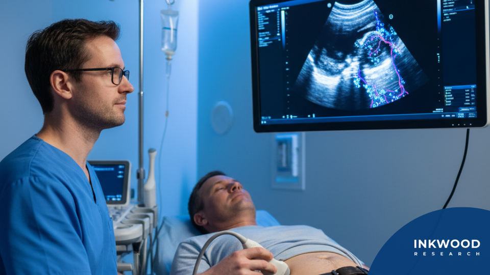
The global contrast enhanced ultrasound market is expanding steadily, driven by aging populations, rising chronic disease burdens, and growing demand for radiation-free diagnostic alternatives to CT and MRI.
India’s contrast ultrasound market is projected to grow from US$72.95 million in 2026 to US$169.89 million by 2034 at an 11.15% CAGR, one of the highest growth rates globally. Surging NAFLD prevalence, ranging from 9% to 32% of adults according to India’s Ministry of Health, is creating structural demand for safe, repeatable hepatic imaging.
South Korea’s market is growing at a 6.93% CAGR, above the global average. CKD prevalence of over 8.2% among Korean adults drives consistent demand for nephrotoxicity-free renal and vascular imaging, where CEUS achieves 91.1% sensitivity for severe renal artery stenosis detection.
Thailand grows from US$28.14 million in 2026 to US$55.51 million by 2034 at 8.86% CAGR, anchored by liver cancer burden and expanding oncology cardiology applications across public and private hospitals.
The United States remains the largest market globally, growing from US$729.85 million to US$1,088.15 million by 2034 at 5.12% CAGR. FDA approval complexity and CPT code gaps for noncardiac applications continue shaping adoption pace despite strong underlying clinical demand.
Key Takeaways
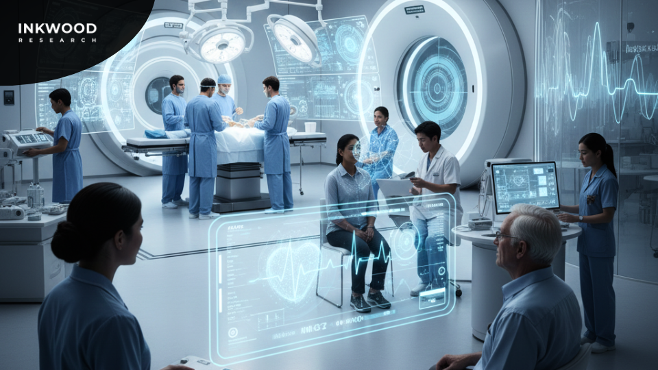
- The global imaging medical devices market reached $18.7 billion in 2026, projected to grow toward $28.2 billion by 2034 at 5.23% CAGR.
- Poland invested approximately $400 million by 2026 in AI-driven medical technologies, revolutionizing mobile imaging services through government-funded infrastructure.
- Medicare’s 2026 dual conversion factor implementation created significant reimbursement challenges for U.S. mobile imaging providers with a controversial -2.5% efficiency adjustment.
- South Korea leads globally in AI-powered retinal disease detection, with systems identifying subtle structural changes predicting future disease development before symptoms appear.
- Indian companies like Forus Health and Remidio revolutionized retinal imaging through affordable, portable solutions achieving 95% diagnostic accuracy while reaching 10 million lives across 50 countries.
- China’s aggressive shift toward home-based cardiac monitoring addresses needs of 290 million people aged 60+, with manufacturers like Lepu Medical and Mindray developing comprehensive telemedicine ecosystems.
- India’s refurbished ECG market evolved from gray market into legitimate industry offering Tier-II and Tier-III hospitals premium technology at 40-50% discounts.
- Optical imaging market reached $3.1 billion in 2026, projected toward $7.5 billion by 2036, reflecting transition from niche applications to mainstream adoption across specialties.
- Dental Support Organizations transformed equipment purchasing patterns, with single decisions by DSOs like Heartland Dental influencing markets more than thousands of individual practice purchases.
- American dental AI systems demonstrate 43% improvement in cavity detection rates, with FDA’s streamlined Class II approval pathway enabling 12-18 month market entry.
- Hybrid operating rooms cost $3-5 million compared to $1-2 million for traditional ORs, yet adoption accelerates globally as certain procedures cannot be performed optimally without integrated imaging capabilities.
- Thailand’s medical tourism boom drove $4 billion in healthcare revenue from 2.5 million annual patients, with hospitals like Bumrungrad International investing heavily in hybrid ORs as marketing differentiators.
- Germany’s systematic approach to hybrid OR adoption emphasizes comprehensive procedural integration, with manufacturers like Siemens Healthineers benefiting from close clinical collaborations accelerating technology refinement.
- Japan’s aging demographics (29% over age 65, projected 38% by 2060) create sustained demand for diagnostic imaging, with portable and AI-powered systems addressing elderly mobility limitations.
- France’s measured adoption pattern reflects healthcare culture demanding rigorous clinical evidence, with COVID-19 accelerating telemedicine acceptance and handheld camera deployment for screening programs and satellite clinics.
Conclusion
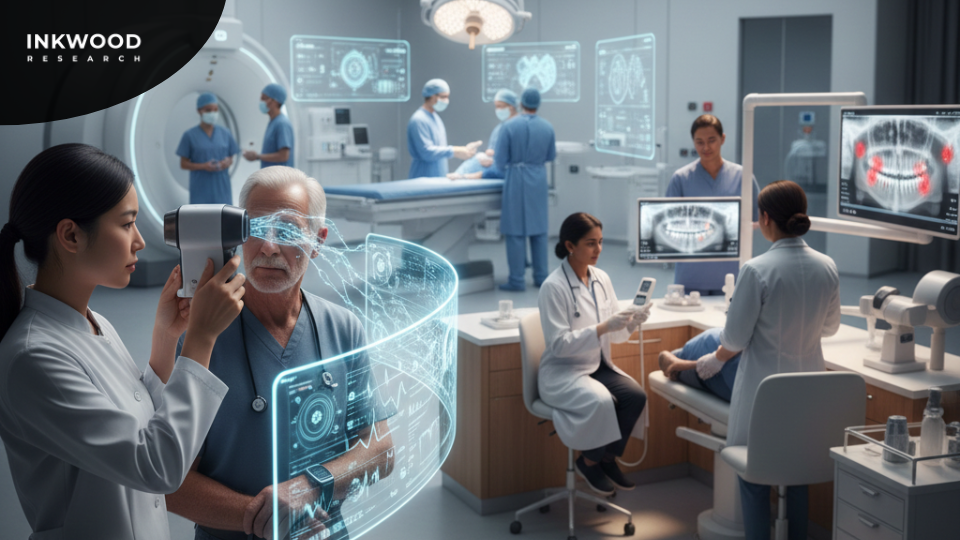
The medical imaging landscape is experiencing a remarkable transformation driven by technological convergence and regional innovation. What’s particularly fascinating is how different nations are carving unique pathways toward the same goal—improving healthcare access and diagnostic accuracy. South Korea’s AI-first approach contrasts beautifully with India’s focus on affordable portability, while China’s ecosystem thinking demonstrates how comprehensive health system adaptation matters as much as the technology itself.
Moreover, the lines between consumer electronics and medical devices continue blurring. Apple Watch detecting atrial fibrillation episodes that cardiologists initially dismissed as “wellness toys” exemplifies how innovation often arrives from unexpected directions. Meanwhile, hybrid operating rooms transition from specialty capabilities toward mainstream infrastructure, dental AI revolutionizes diagnostic accuracy, and optical imaging creates entirely new clinical possibilities across multiple specialties.
Perhaps most encouraging is how these advances address genuine healthcare access gaps rather than simply adding expensive capabilities for wealthy populations. India’s refurbished equipment markets, Poland’s government-funded mobile imaging, and portable retinal cameras reaching rural communities all demonstrate technology’s democratizing potential. As we look toward 2034 and beyond, the medical imaging market’s projected growth reflects not just economic opportunity but genuinely improved patient outcomes worldwide through smarter, more accessible diagnostic capabilities.